
The Wave - The characteristics of an EEG
Welcome back!
I see you’ve got a hang of the basics of nerve impulse lingo from our previous article(link). Let’s proceed to the next aspect - the EEG waves.
By now, I would expect you to have asked one question to yourself. There is so much talk of neurons firing, and this and that. The brain is almost basically made up of neurons, right?
So, which neurons are we talking about when we say ‘neurons are firing’?
It’s a very valid question. As you know, the brain is divided into a multitude of areas, each with its own function. So when we keep an electrode in the occipital lobe, we are essentially measuring the activity of the neurons in and around that area. Neurologists have determined specific areas where we need to place electrodes to ensure we end up with a precise and accurate EEG. If they hadn’t, like you suspected keeping electrodes in random places on the brain will pick up random electrical signals from neurons in that area and we wouldn’t be able to assess anything properly.
As we all know, our brains are constantly in activity. Even while we rest, the neurons in our brains are busy sending impulses. So even if we take an EEG for a person who is sleeping peacefully, there is still excitation at the level of the neurons. What matters here is the level of excitation. Obviously, the level of excitation is less while we’re sleeping- nevertheless, it's still there.
So now we know that the electrical impulses recorded from the brain are constant and that they vary based on our state of mind. So how do we differentiate these waves from one another? Well, in order to describe any wave we need to know two features of it: the strength and the frequency (cycles per second). The strength of current, or the voltage range of the neurons is from 0 to 200 microvolts- yes, you read it right correctly. Microvolts. The frequency ranges from 1 to 50 times per second. It’s all the same content you learned in physics - just applied at the micro-cellular level.
Based on these two characteristics the commonly seen brain waves are divided into: Alpha, beta, theta, delta.
Let’s look at this table to help us differentiate between these waves in terms of the voltage and frequency.
|
WAVE |
VOLTAGE |
FREQUENCY |
|
α(alpha) |
50 mV |
8-13 Hz |
|
β(beta) |
< 50 mV |
14-30 Hz |
|
Θ(theta) |
50 mV |
4-7 Hz |
|
Δ(delta) |
100 to 200 mV |
< 3.5 Hz |
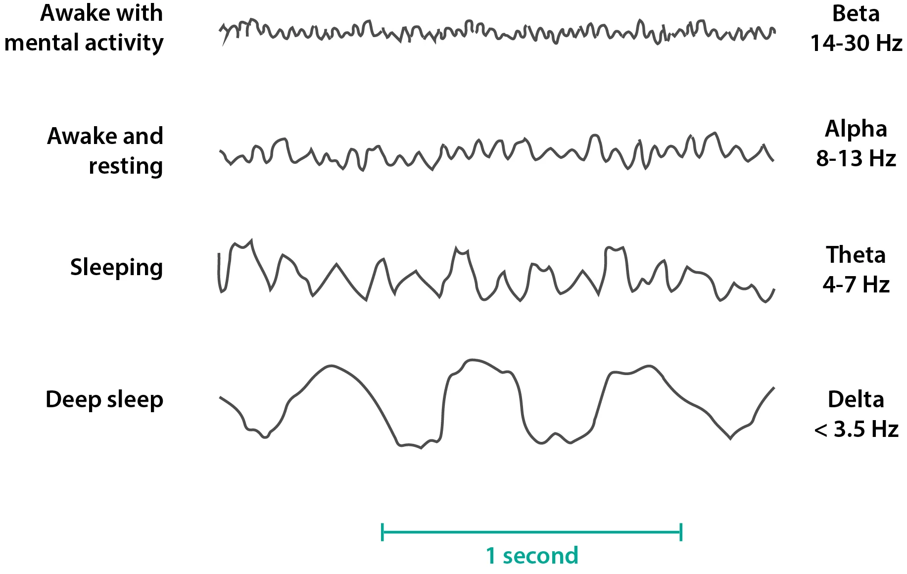
As you can see, alpha waves are similar to theta waves, but they have a higher frequency. Beta waves are really low in voltage but they have the highest frequency. Theta and delta waves are similar to alpha waves but they are really slow. Their frequency is almost half of that of the alpha waves.
Now, here comes the slightly tricky part. Remembering which waves are seen where. Like I said before, the electrodes are placed in specific places, which helps us standardize EEGs and makes their interpretation much easier.
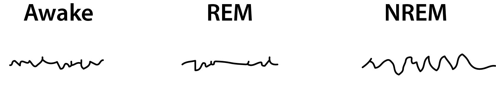
When you’re sleeping, you’re in a state of calm. Your thoughts are free to roam wherever and there is no focus of thought. You aren’t thinking of anything specific, but your brain is just wandering around, thinking its own thoughts (and giving you really bizarre dreams sometimes!) You can correlate this with theta and delta waves. Frequency low but voltage somewhat high. Not many thoughts but wide ranging thoughts nevertheless.
When you’re closing your eyes, your visual stimulus is lost, so you are confined to your other senses and your thoughts. However, when you’re awake and your eyes are open, the capacity to send impulse is increased. Think of it this way- eyes closed: less distractions, eyes open: a lot of distractions, a lot of things to think about, a lot of stimulus and therefore- the frequency of firing is increasing. While awake, there is so much information to be processed that the brain has to manage by increasing the number of impulses sent rather than the strength of impulses. Got it?
Take a look at this EEG:
Let’s test ourselves at this point. Now that you’ve got an understanding of the way the waves are formed, let’s try to figure out where each type of wave is seen.
We’ve seen that delta and theta waves are seen in sleep
What about in states of stress?
Did you guess theta waves? If so, you’re right. If you didn’t, (or if you’re wondering why your random guess is correct), stress causes increased total firing of the neurons, but decreased frequency. Simply put, it’s like this- the more the frequency of firing (to a limit, of course) you can interpret the reading as, the brain is able to receive and send information at a normal pace. When you’re stressed, your output is high, but you find it difficult to focus on more than one thing. Therefore, the big but slow waves.
What about in organic brain diseases?
For similar reasons as stated above, theta and delta waves predominate.
So these are the normal waves of the EEG. The basic point here is that the wave information is collected from specific predetermined points on the scalp. These points are representative of vital areas in the brain. The electrical information is then interpreted based on the frequency and the amplitude of the waves to give us an understanding of the type of pathology and the location of the pathology.
In our next article we will discuss the parts of the brain from where these waves arise.
Author: Shruthi Sivakumar
Sources and citations
Hall, John E. “States of Brain Activity - Sleep, Brain Waves, Epilepsy, Psychoses.” Guyton and Hall Textbook of Medical Physiology, 12th ed., Elsevier Saunders, 2011, pp. 723–726.